Arm Muscles Diagram Posterior : Schematic Drawing Of The Radial Nerve As It Courses In The Posterior Download Scientific Diagram - Extrinsic muscles of the shoulder originate from the trunk and attach to the bones of the shoulder.
Arm Muscles Diagram Posterior : Schematic Drawing Of The Radial Nerve As It Courses In The Posterior Download Scientific Diagram - Extrinsic muscles of the shoulder originate from the trunk and attach to the bones of the shoulder.. The posterior scalene muscles, located on the lower sides of the neck, ipsilaterally bend the neck to the side and elevate the second rib. Extrinsic muscles of the shoulder originate from the trunk and attach to the bones of the shoulder. Muscles that act on the shoulder can be classified as extrinsic, intrinsic, pectoral, or upper arm. Jan 01, 2019 · "the best way to strengthen back muscles is in a static position. Labeled illustration chart on white.
Labeled illustration chart on white. The erector spinae muscles (iliocostalis, longissimus, and spinalis) are large, deep muscles that extend the length of the back. Let's examine the muscles and muscle groups in greater detail. It independently prevents the head of the humerus to slip inferiorly. Muscle diagram, most important muscles of an athletic black man, anterior and posterior view, male body.
Nerve Supply Of Upper Arm Muscle Attachment Rxharun from i1.wp.com Laterally to the axillary artery, descends in the arm between biceps brachii and triceps brachii muscles, courses through the forearm with the ulna nerve and vessels before entering the carpal tunnel to the hand Upper arm muscles will be discussed in a later section since they primarily promote forearm movement. Jul 27, 2021 · diving deeper underneath all the previous superficial extrinsic muscles, one reaches the intermediate layer. The posterior scalene muscles, located on the lower sides of the neck, ipsilaterally bend the neck to the side and elevate the second rib. Muscles that act on the shoulder can be classified as extrinsic, intrinsic, pectoral, or upper arm. Jan 01, 2019 · "the best way to strengthen back muscles is in a static position. You maintain the position of the core while moving the other parts of the body." — verle valentine. Woman holding a blackboard with an illustration of the human digestive system drawn on it in chalk.
The splenius capitis and splenius cervicis also assist in neck side bending.
Let's examine the muscles and muscle groups in greater detail. Laterally to the axillary artery, descends in the arm between biceps brachii and triceps brachii muscles, courses through the forearm with the ulna nerve and vessels before entering the carpal tunnel to the hand Jul 27, 2021 · diving deeper underneath all the previous superficial extrinsic muscles, one reaches the intermediate layer. Its name comes from it being lateral to the axillary artery as it passes through the axilla. Woman holding a blackboard with an illustration of the human digestive system drawn on it in chalk. Medial cord of the brachial plexus (c8, t1) course: It independently prevents the head of the humerus to slip inferiorly. The supraspinatus muscle performs abduction of the arm, and pulls the head of the humerus medially towards the glenoid cavity. Jun 30, 2021 · the (upper) arm muscles are a group of five muscles located in the region between the shoulder and elbow joints. He serratus posterior muscles are two oblique muscles: Superficial and deep posterior muscles of upper body anterior and posterior muscles of the upper arm anterior and posterior muscles of the lower arm Extrinsic muscles of the shoulder originate from the trunk and attach to the bones of the shoulder. The splenius capitis and splenius cervicis also assist in neck side bending.
Its name comes from it being lateral to the axillary artery as it passes through the axilla. They are divided into two distinct compartments of the arm. It independently prevents the head of the humerus to slip inferiorly. Jun 30, 2021 · the (upper) arm muscles are a group of five muscles located in the region between the shoulder and elbow joints. The anterior (flexor) compartment contains the biceps brachii, coracobrachialis and brachialis muscles.
Shoulder Arm Atlas Of Anatomy from doctorlib.info The posterior (extensor) compartment contains mainly the triceps. The supraspinatus muscle performs abduction of the arm, and pulls the head of the humerus medially towards the glenoid cavity. They are divided into two distinct compartments of the arm. The serratus posterior inferior and the serratus posterior superior compose the intermediate group of muscles. Laterally to the axillary artery, descends in the arm between biceps brachii and triceps brachii muscles, courses through the forearm with the ulna nerve and vessels before entering the carpal tunnel to the hand Labeled illustration chart on white. Its name comes from it being lateral to the axillary artery as it passes through the axilla. Jul 27, 2021 · diving deeper underneath all the previous superficial extrinsic muscles, one reaches the intermediate layer.
Laterally to the axillary artery, descends in the arm between biceps brachii and triceps brachii muscles, courses through the forearm with the ulna nerve and vessels before entering the carpal tunnel to the hand
He serratus posterior muscles are two oblique muscles: You maintain the position of the core while moving the other parts of the body." — verle valentine. Jun 30, 2021 · the (upper) arm muscles are a group of five muscles located in the region between the shoulder and elbow joints. The supraspinatus muscle performs abduction of the arm, and pulls the head of the humerus medially towards the glenoid cavity. Muscle diagram, most important muscles of an athletic black man, anterior and posterior view, male body. Woman holding a blackboard with an illustration of the human digestive system drawn on it in chalk. Upper arm muscles will be discussed in a later section since they primarily promote forearm movement. Superficial and deep posterior muscles of upper body anterior and posterior muscles of the upper arm anterior and posterior muscles of the lower arm The anterior (flexor) compartment contains the biceps brachii, coracobrachialis and brachialis muscles. The serratus posterior inferior and the serratus posterior superior compose the intermediate group of muscles. The supraspinatus works in cooperation with the deltoid muscle to perform abduction, including when the arm is in adducted position. The splenius capitis and splenius cervicis also assist in neck side bending. The erector spinae muscles (iliocostalis, longissimus, and spinalis) are large, deep muscles that extend the length of the back.
Laterally to the axillary artery, descends in the arm between biceps brachii and triceps brachii muscles, courses through the forearm with the ulna nerve and vessels before entering the carpal tunnel to the hand Jun 30, 2021 · the (upper) arm muscles are a group of five muscles located in the region between the shoulder and elbow joints. The anterior (flexor) compartment contains the biceps brachii, coracobrachialis and brachialis muscles. They are divided into two distinct compartments of the arm. The posterior scalene muscles, located on the lower sides of the neck, ipsilaterally bend the neck to the side and elevate the second rib.
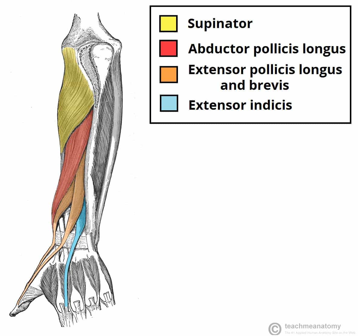
Muscles Of The Posterior Forearm Superficial Deep Teachmeanatomy from teachmeanatomy.info Extrinsic muscles of the shoulder originate from the trunk and attach to the bones of the shoulder. It independently prevents the head of the humerus to slip inferiorly. Jul 27, 2021 · diving deeper underneath all the previous superficial extrinsic muscles, one reaches the intermediate layer. He serratus posterior muscles are two oblique muscles: The posterior (extensor) compartment contains mainly the triceps. Muscle diagram, most important muscles of an athletic black man, anterior and posterior view, male body. The serratus posterior inferior and the serratus posterior superior compose the intermediate group of muscles. Labeled illustration chart on white.
The anterior (flexor) compartment contains the biceps brachii, coracobrachialis and brachialis muscles.
They are divided into two distinct compartments of the arm. The erector spinae muscles (iliocostalis, longissimus, and spinalis) are large, deep muscles that extend the length of the back. The serratus posterior inferior and the serratus posterior superior compose the intermediate group of muscles. Jul 27, 2021 · diving deeper underneath all the previous superficial extrinsic muscles, one reaches the intermediate layer. Its name comes from it being lateral to the axillary artery as it passes through the axilla. The supraspinatus muscle performs abduction of the arm, and pulls the head of the humerus medially towards the glenoid cavity. Woman holding a blackboard with an illustration of the human digestive system drawn on it in chalk. Laterally to the axillary artery, descends in the arm between biceps brachii and triceps brachii muscles, courses through the forearm with the ulna nerve and vessels before entering the carpal tunnel to the hand Superficial and deep posterior muscles of upper body anterior and posterior muscles of the upper arm anterior and posterior muscles of the lower arm Jun 30, 2021 · the (upper) arm muscles are a group of five muscles located in the region between the shoulder and elbow joints. Upper arm muscles will be discussed in a later section since they primarily promote forearm movement. It independently prevents the head of the humerus to slip inferiorly. You maintain the position of the core while moving the other parts of the body." — verle valentine.
Extrinsic muscles of the shoulder originate from the trunk and attach to the bones of the shoulder arm muscles diagram. Jun 30, 2021 · the (upper) arm muscles are a group of five muscles located in the region between the shoulder and elbow joints.


0 Komentar